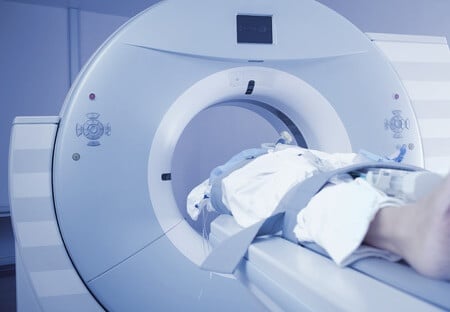
This case involves a patient who developed pulmonary hypertension after using the drug Fenfluramine/phentermine for a one year period. The patient experienced chest pain and severe shortness of breath and was subsequently admitted to the hospital for a workup to rule out thromboembolic disease as a potential cause. The original CT-angiogram was read as having a low index of suspicion for thromboembolic disease. A short time after the patient underwent a perfusion scan and a pulmonary angiogram that revealed chronic embolic disease.
Question(s) For Expert Witness
- 1. Can you determine from chest imaging whether it is likely that the patient had pulmonary hypertension at the time the embolism was diagnosed and if so how does pulmonary hypertension increase the risk of thromboembolic disease?
Expert Witness Response E-000888
Thromboembolic disease leads to pulmonary hypertension. A CT angio is good at picking up an acute PE, but chronic disease is best picked up on a V/Q or angiogram. Enlargement of the pulmonary vessels on CT could be suggestive, but not diagnostic of pulmonary hypertension. Chronic thromboembolic disease causes pulmonary hypertension. Pulmonary hypertension can worsen thromboembolic disease, in my opinion, mostly indirectly and Phen-Fen has been known to cause Pulmonary Hypertension. To be clear a pulmonary angiogram is the gold standard method of diagnosing chronic thrombosis.
Contact this expert witness
Related Posts
This case involves a stroke patient who underwent an endoscopic PEG tube placement and deteriorated shortly thereafter. A CT scan showed significant evidence of pneumo-peritoneum, likely related to gastrostomy tube placement. An exploratory laparotomy was performed the same day and…
This case involves a 5-year-old female who presented to the ER with abdominal pain. She had a past medical history significant for developmental delay and required an intravenous total parenteral nutrition feeding tube placement (TPN) several months prior. The patient…